

Research
Research outline
We have been developing atomic force microscopy (AFM), one of the atomic scale measurement techniques, and using it for research in various academic and industrial fields. AFM is a measurement technique that enables observation of a surface structure of an object at atomic resolution by precisely tracing the surface with a sharp probe (figure below). Among several AFM operation modes, frequency modulation AFM (FM-AFM) has the highest resolution. Professor Fukuma has realized the world’s first atomic resolution FM-AFM observation in liquid. Based on this technical basis, we are working on developing and applying advanced in-liquid AFM measurement techniques.
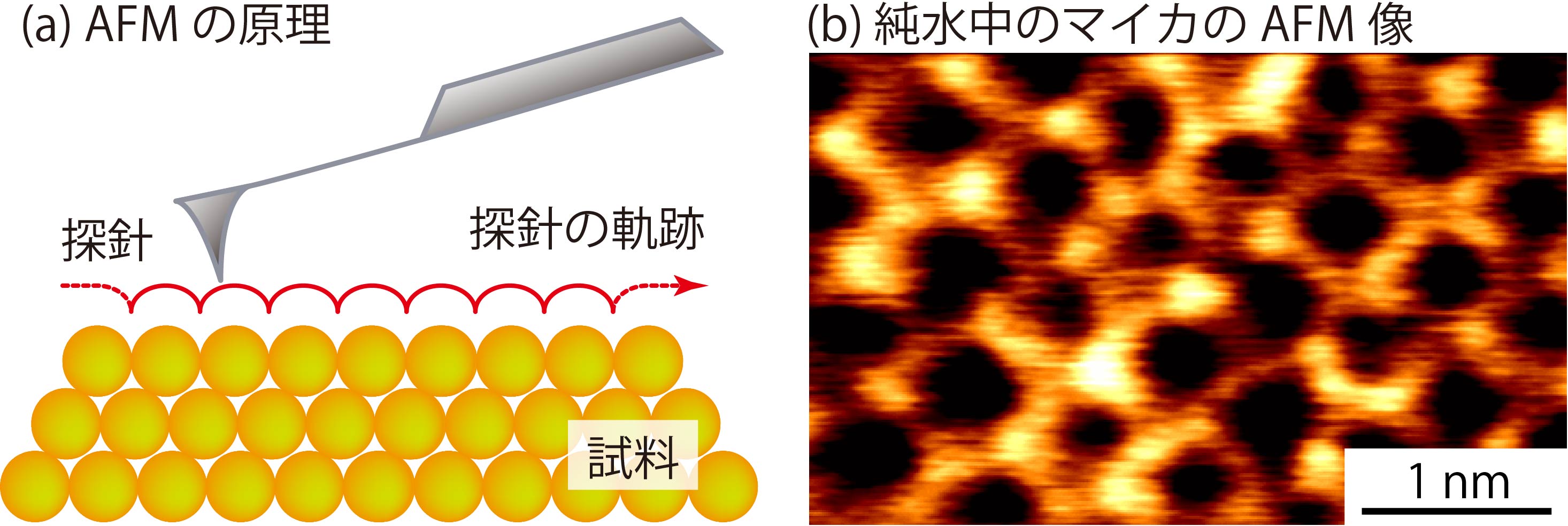
Atomic -resolution imaging observation by in-liquid FM-AFM for the first time in the world [1]
Instrumentation of AFM
【Design and development of self-made AFM from scratch】
There are many research groups around the worldworldwide working on the development of AFM techniques, but most of them are working on modifications based on commercially available AFMs. On the other hand, in our laboratory, we design and develop our own AFM from scratch; it enables us to develop a new measurement technique and disruptively improve basic performance exponentially, which cannot be realized by simply modifying commercially availableready-made instrumentsproducts. We also actively apply for patents on our developed technique and transfer the technique to domestic and overseas AFM manufacturers.
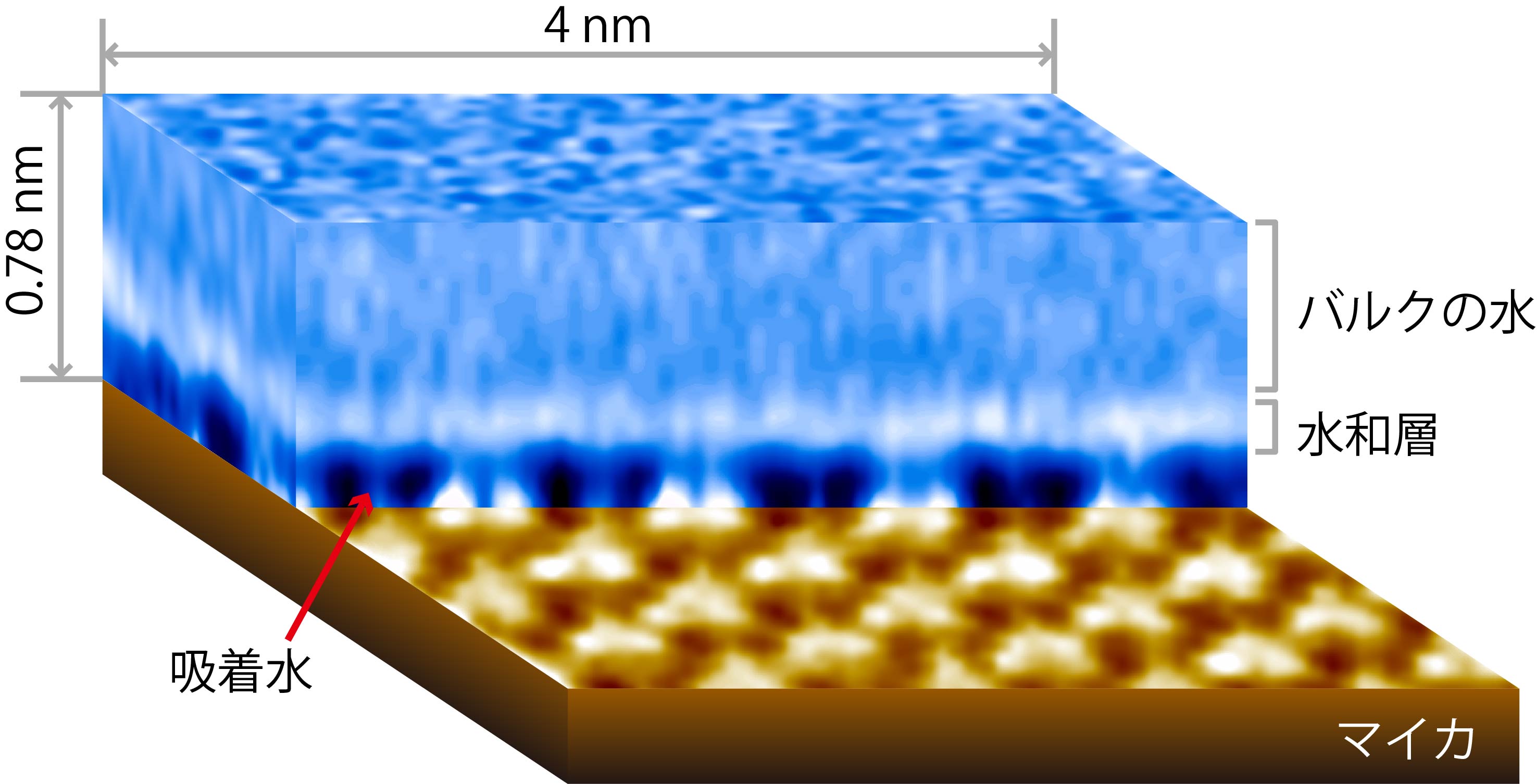
Sub-nanoscale visualization of a 3D hydration structure on a solid-liquid interface for the first time in the world [2]
【Development of novel measurement methods】
We are developing novel measurement methods based on the in-liquid AFM technique. For example, we have developed a technique that can measure 3D force distribution near the sample surface and succeeded in directly observing the 3D distribution of a hydrated layer and adsorbed water for the first time in the world.
Hydration is a phenomenon that commonly occurs on the surface of materials around us and is deeply involved in the function of materials and devices. There had been no method to observe hydration phenomena on a molecular scale; however, we have developed this novel method that enables the observation for the first time. This method is expected to be useful for research and development in various academic and industrial fields.
【Dramatic improvements in basic performance】
We are developing new techniques that dramatically improve the basic performance of in-liquid AFM, such as sensitivity and speed. For example, we have significantly improved the force sensitivity by downsizing the sensor called “cantilever.”
This improvement in the basic performance has significant impactsripple effects. First, the improved sensitivity increases the reproducibility and reliability of AFM measurements. It also enables observation of the 3D hydration structure with higher accuracy than before and detailed comparison ofwith the results with those of theoretical simulations in detail. This is a significant advance in the quantitative interpretation of measurement results. Furthermore, the improved sensitivity allows us to perform atomic-resolution observations faster than before. We have improved FM-are working on the development of AFM imaging speed with the aim of speeding up the observation time from ~about 1 min/frame one minute per frame to ~1 sec/frameabout one second per frame.
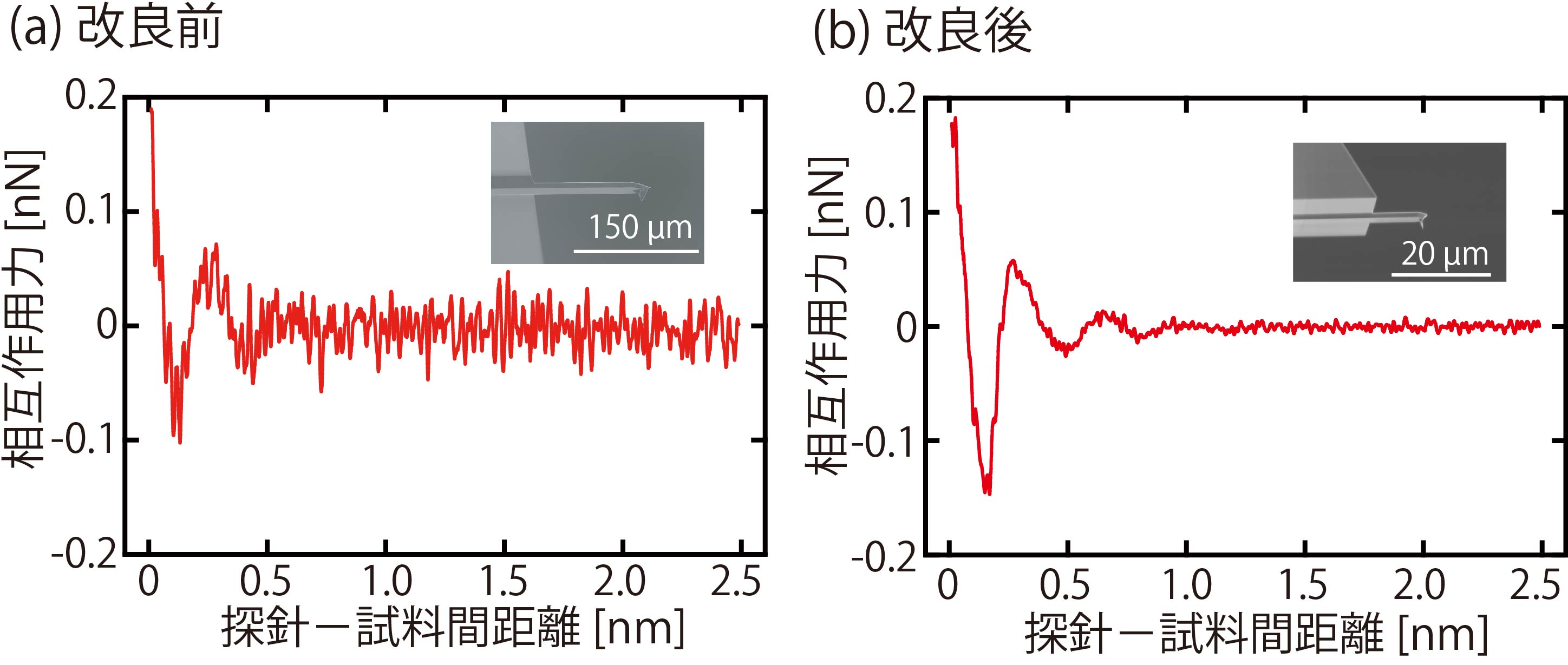
Force-distance curves obtained on a mica surface in water. The force sensitivity is greatly improved by reducing the size of the cantilever [3]
Applied research
【Application of cutting-edge AFM technique to a wide range of research fields】
Although measurement and analysis techniques such as AFM are not something we use directly in our daily lives, they are widely used in the research and development of a variety of products related to our daily lives. AFMs are often installed in analysis centers of universities and R&D departments of companies. Therefore, the advancement of AFM application techniques will benefit wide-ranging academic and industrial fields. While AFMs are used in various fields, the most important feature of our research is that we are trying to realize measurements that have never been achieved before by using the globally advanced AFM techniques that have just been developed. This is possible only because our laboratory conducts the entire process from development to application. Until now, the atomic-level AFM measurement technique has been used exclusively for basic academic research on relatively simple samples and phenomena in the fields of surface physics and chemistry. In contrast, we are challenging not only research in the field of physics and chemistry, but also observation of cells and proteins in the field of biophysics, and evaluation of semiconductor interconnections and plant materials in the industrial field. In this way, we are challenging diverse issues, from basic academic research to the development of industrial technology, using atomic-level measurement techniques. It makes our research peerless in the world. Through these applicationsis applied research, we can find quickly identify measurement needs as early as possible and feedback themapply the needs to the development of next-generation techniques and equipment, which allows us to conduct world-leading research and development.
【Application to biomolecular measurements】
In addition to AFM, transmission electron microscopy (TEM) and scanning tunneling microscopy (STM) are other atomic-level measurement techniques. However, AFM is the only technique that enables imaging insulating materials in liquid with atomic resolution. This feature is extremely useful for research in biology, where biomolecules with poor electrical conductivity need to be analyzed in liquid. Therefore, we are developing techniques applicable to high-resolution in-liquid AFM imaging of biological systems.
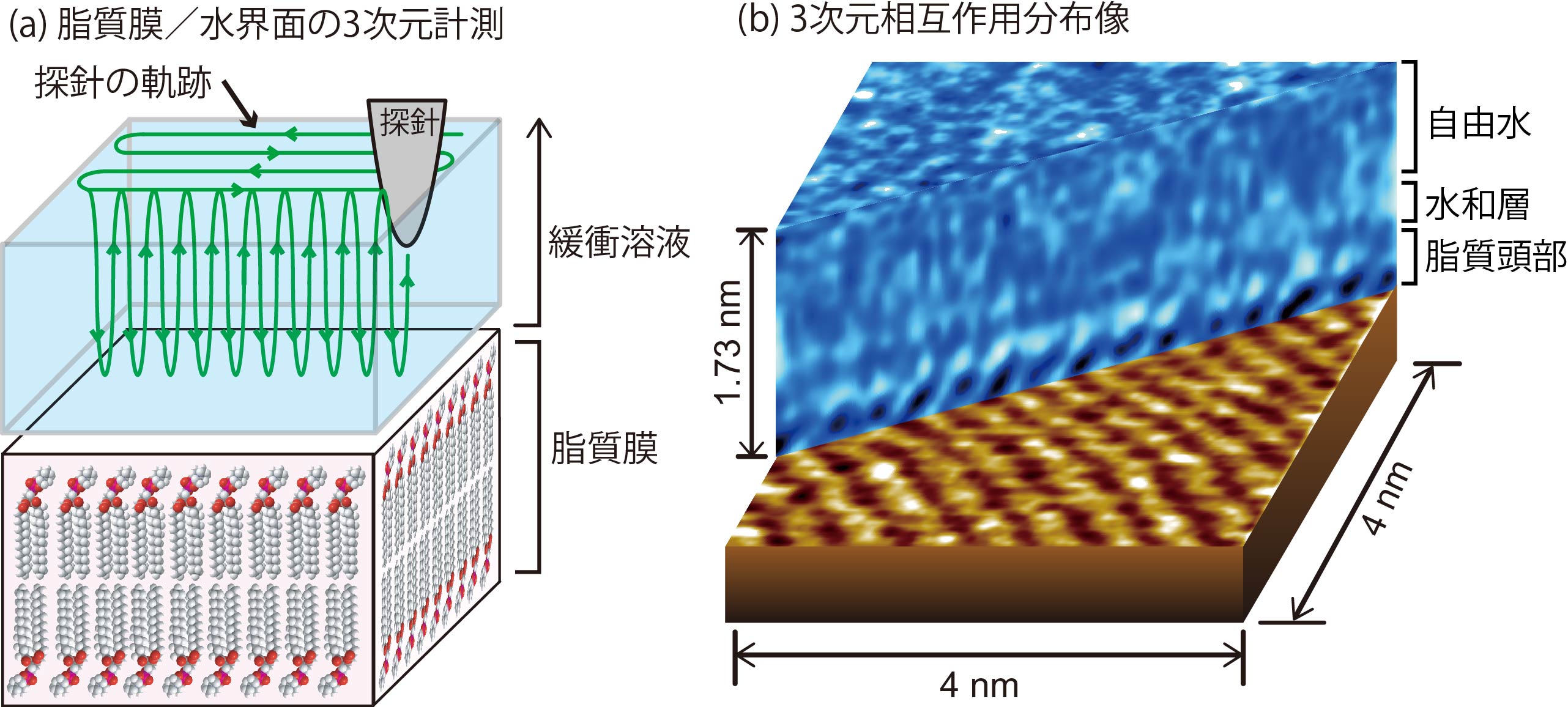
Visualization of the three-dimensional distribution of hydration layer and lipid head groups at the lipid membrane/water interface [4]
For biological applications, we are working on measuring biomolecular systems with different dimensions, such as lipid membranes, protein aggregates, and cells. For example, we have measured the 3D force distribution at the interface between lipid membranes and water and found that the obtained images reflect not only the hydration layer of the lipid membrane surface but also the 3D fluctuating structure of the lipid head groups that compose the lipid membrane surface. Although lipid membranes have been widely studied as a model of cell membranes, our in-liquid AFM is the first to visualize the hydration and fluctuating structures on their surfaces at the molecular level. This research result showed for the first time that 3D measurement can visualize not only the hydration structure but also the surface fluctuating structure.
In addition, we have succeeded in directly observing α- helices and C-termini in liquid at the subnanoscale for the first time in the world. The α- helix and C-terminus exist on the surface of a tubulin complex. Conventional AFM technique can visualize individual tubulin molecules, but not the fine structure within the molecules. We have enabled the observation of the fine structures by making full use of our originally developed high-resolution AFM.
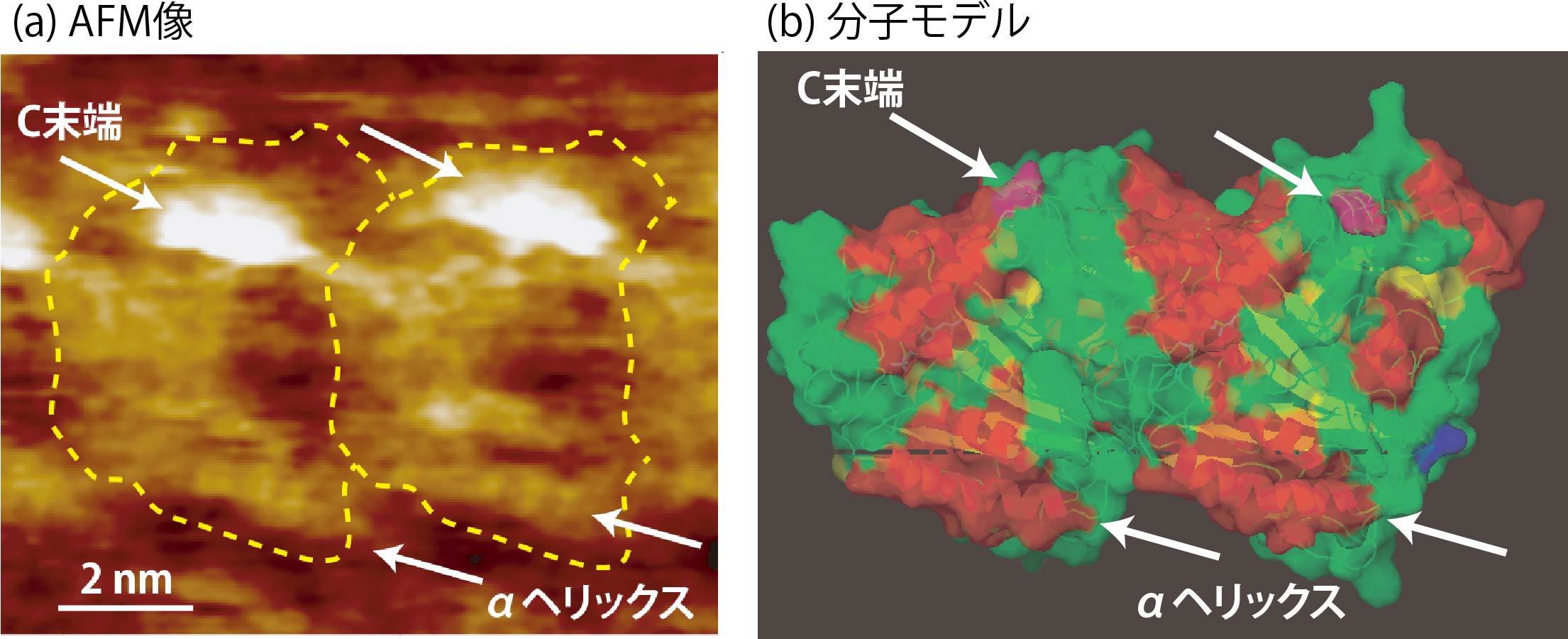
Subnanoscale visualization of alpha- helices and C-termini on a surface of tubulin molecules [5]
In living organisms, the surface of a protein complex is an important place where inter-molecular interactions in physiological solutions occur, and these interactions have a significant impact on the mechanisms of various biological functions. Therefore, if the fine structures of protein surfaces can be determined in detail using in-liquid AFM, it should lead to a better understanding of the effects of molecular interactions on biological functions.
【Applying AFM to other research fields】
At the beginning of our laboratory, we mainly focused on biophysics research. Since ~2010, we also started to apply our techniques to the studies in interface sciences and electrochemistry. The number of such research subjects has gradually increased, and now they account for more than half of our AFM applications. In physical chemistry, we investigate mechanisms of hydration, crystal growth, and dissolution on the surface of mineral crystals such as calcite (CaCO3) and fluorite (CaF2) at the atomic level. In addition, we investigate the effects of molecular fluctuations on surface hydrophilicity and molecular adsorption properties at the interface between water and surfaces composed of highly fluctuating molecular structures such as polymeric materials.
Our research for industrial technology is mainly carried out in collaboration with private companies from various fields, aiming to use our findings for their research and development.
References
[1] T. Fukuma, K. Kobayashi, K. Matsushige and H. Yamada, “True Atomic Resolution in Liquid by Frequency-Modulation Atomic Force Microscopy” Appl. Phys. Lett. 87 (2005) 034101 (3 pages).
[2] T. Fukuma, Y. Ueda, S. Yoshioka, H. Asakawa, “Atomic-Scale Distribution of Water Molecules at the Mica-Water Interface Visualized by Three-Dimensional Scanning Force Microscopy” Phys. Rev. Lett. 104 (2010) 016101 (4 pages).
[3] T. Fukuma, K. Onishi, N. Kobayashi, A. Matsuki, H. Asakawa, “Atomic-resolution imaging in liquid by frequency modulation atomic force microscopy using small cantilevers with megahertz-order resonance frequencies” Nanotechnology 23 (2012) 135706 (12 pages).
[4] H. Asakawa, S. Yoshioka, K. Nishimura, T. Fukuma “Spatial Distribution of Lipid Headgroups and Water Molecules at membrane/Water Interfaces Visualized by Three-Dimensional Scanning Force Microscopy” ACS NANO 6 (2012) 9013–9020.
[5] H. Asakawa, K. Ikegami, M. Setou, N. Watanabe, M. Tsukada, T. Fukuma,“Submolecular-Scale Imaging of α-Helices and C-Terminal Domains of Tubulins by Frequency Modulation Atomic Force Microscopy in Liquid” Biophys. J. 101 (2011) 1270-1276.